Interpreting Tree Decay
By David Humphries, September 7, 2017 in General chat
Can early stage decay caused by Kretzschmaria deusta be detected & differentiated from other types of decay using a micro-drill?
David Humphries and Alasdair Nicoll
City of London Corporation Arborists – Hampstead Heath, North London Open Spaces
There have been suggestions that some decay detection equipment like the IML Resi PD400 may struggle to detect the early stages of decay caused by fungi species like Kretzschmaria deusta, which are known to exhibit a soft rot mode of decay, 1 sometimes referred to as a ‘facilitative rot’ mode, meaning it’s establishing conditions for the progress of decay. We decided to test this theory by comparing a cross-section of a failed lime tree against micro-drill readings taken from different points around the circumference through sound, dysfunctional and decayed wood.
For the tests described in this article we have used the IML Resi PD400. It is a micro drill used for tree & utility pole inspection that tests for resistance through a given wood volume. A consistent force is applied and measured. As the probe penetrates the volume the difference between denser and less dense wood is measured which then indicates wood condition.
#jscode#
The IML Resi PD series has the option of 5 needle speed settings that govern rpm & rate of feed. Different speed enables the probe to pass through differing densities of the wood volume. It is worth considering that amplitude (height of the reading) can be read higher up the Y axis based upon the speed setting of the needle. Amplitude is not a literal reading of the wood quality it is simply a change in resistance and should not be taken as a signature for any specific decay species and/or type.
Main types of decay
Soft rot/facilitative rot – Wood initially becomes brittle due to cellulose degradation. This often results in ceramic brittle fracture of affected parts, should they fail.
Example: Kretzschmaria deusta 2
Brown rot – This rot type primarily degrades cellulose and hemicellulose, leaving the lignin intact. Wood shrinks noticeably in volume, sometimes cracking, becomes lighter and more prone to fracture across the wood grain.
Example: Laetiporus sulphureus 2
White rot – This type of rot can degrade all three major constituents of wood: cellulose, hemicellulose & lignin. Two different types of white rot are commonly reported:
Simultaneous white rot – all three constituents are broken down at similar rates.
E.g. Fomes fomentarius 2
Selective white rot – lignin & hemicellulose are broken down initially, leaving cellulose relatively unaffected. Wood becomes soft, lighter in weight and colour and often much more stringy/fibrous. E.g. Ganoderma sp 2
in this article we look predominantly at a test case involving early and late stage decay caused by K. deusta within the same tree & how they can be identified across a set of IML Resi PD400 readings.
Context
During zonal inspection in 2012 fruit bodies of K.deusta were first noted on a mature common lime tree (Tilia x europaea) growing beside a well used pedestrian avenue in north London.
The canopy was reduced to balance the weight of tree away from the footpath at that time. Cracks were then identified on the trees buttresses on the path side during the winter of 2015.
The following observations outlined in this article have been produced in the field testing only a narrow range of specimens and not under laboratory conditions, measurements are approximate. The IML Resi PD400 readings are consistent with examples taken on other cross sections and standing trees.
Observations of decay
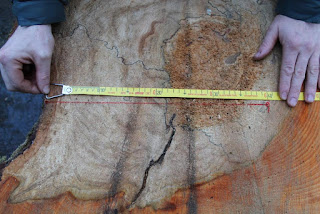
After the failure of the subject tree we had the opportunity to test a decayed section of the tree by taking a 4cm thick cross section of the lower stem and buttresses.
Readings were taken by drilling through five locations using the IML Resi PD400 to observe the mechanical differences across between intact (sound) wood, early stage decay (including
reaction zone) and late stage decay that had the consistency of a severe white rot. In our opinion this may be late stage K.deusta decay or a separate white rot species.
Figures 1 and 2 above; The lime, showing separation cracks at ground level and fruiting bodies of K. deusta
Figures 1 and 2 above; Post failure images showing where the lower buttresses have fractured.
Figures 5 and 6 above; A 4cm thick cross section of the lower stem and buttresses.
Test A
A 40 cm IML Resi reading (to be read from right to left) from the outer bark through the early stage decay caused by K. deusta in the sap wood then in to the late stage decay in the centre of the section and then back into early stage decay. (Graph 1 to be read from right to left)
Interpretation
The Test A reading (Graph 1) first shows 3 cm of bark followed by irregular rising peaks and troughs as the drill passed through the early stage decay between approximately 3 & 23.5 cm. This can be seen accentuated by the higher green reading (needle speed torque resistance)
There is then a significant drop off in the reading from 23.5 cm where the needle penetrates the late stage decay for another 14 cm and then continues into the early stage decay for the last 3 cm of the reading & can be seen as a rise in the graph.
Both resistances, torque (green) and penetrating (blue) can be seen as higher across the graph within the early stage decay zone than it can in the late stage decay area due to there being increased resistance to the drill.


Test B
A 40 cm reading was taken as a control drilling through an intact part of the cross section which is functional wood (Figure 8 and Graph 2)
Interpretation
This test was taken to provide a ‘normal’ reading where there was no decay in the cross section. The green & blue readings show a consistent resistance to penetration (blue) and consistent slight increase in resistance to torque (green) due to frictional drag along the drill bit.
(The drop off at the end is due to the needle exiting the cross section underneath)
Test C
A 40 cm IML Resi PD reading taken drilling through outer functional sapwood, then through the early stage decay & in to late stage decay.
Interpretation
Test C is a combination between tests A & B, as it shows both intact functional wood between 1-13 cm, early stage/dysfunction between 13 – 26 cm & late stage decay from 26 cm. The peak at 37 cm is a pocket of significantly denser wood within the more decayed part of the section.
Test D
40 cm IML Resi PD400 reading taken to attempt to observe if there was any noticeable effect on the reading from the presence of pseudosclerotial plates (black demarcation lines) within the cross section.
Interpretation
The above reading appears to suggest that the pseudosclerotial plates could possibly be differentiated as there is a peak at 16 cm where the first black line shows at the crossing point between two red lines labelled C and D on the cross section.


Test E
The following is a shorter (15 cm) IML Resi PD400 reading taken to assess a noticeable reaction zone.
Interpretation
Beyond the functional sapwood where the graph (below) shows a consistent level reading between 3-7 cm, there is then what we perceive to be a definite (green coloured) reaction zone between 7- 8.5 cm which seems to show as more erratic peaking than the functional wood immediately to the right.
The above image appears to show a different level of moisture content in the span of the reaction zone in between the adjacent wet sound and dried decayed wood sections. As found by Pearce (2000)3 This would indicate the changing from functional, through dysfunctional then into decayed wood.
The following example and reading is from a different fallen lime also with evident fruiting bodies of K. deusta at its base, which again appears to show the reaction zone between early stage decay and intact sapwood.
Both torque (green) and penetration (blue) increase in resistance between 4 -5.5 cm before going into the decayed section.


Test F
Figure 12 and Graph 6 show a different fallen lime, also with evident fruiting bodies of K. deusta at its base. The short cm reading again appears to show the reaction zone between early stage decay and intact sapwood. Both torque (green) and penetration (blue) increase in resistance between 4cm and 5.5cm before going into the decayed section.
Figures13 and 14: (above) a standing lime infected on one side by K.deusta was the subject of tests G and H
Test G and H
The next example is a standing lime infected on one side by K. deusta. Which was drilled to show that the decay is detectable without seeing it through a cross section.
The first graph (on the left) reads through the decayed portion of the trunk clearly evident at the outer edge of the stem near to fruiting bodies. The second graph (on the right) was taken on the opposite side of the trunk through functional wood and then into decayed portion of the trunk.
Interpretation
The left graph shows significantly decayed wood through the entire reading whilst the graph on the right shows functional wood until approximately 14.5 cm where the early stage decay extends to.


Visual comparison of other decay types
Here we look at observations of the brown rot caused by Laetiporus sulphureus within oak with the associated IML Resi PD400 reading.
Brown rot Test I
Interpretation
Graph 9 shows functional intact wood from 2 – 15 cm, then the drops off indicating significant brown rot with almost negligible resistance (blue) before penetrating sound wood again. There is some shaft resistance indicated by the blue penetrating force rather than dropping to zero level. If this was early stage decay of K. deusta, this section of the graph (between 17 cm – 28 cm). would show as a higher and more erratic reading of penetration force.
White Rot Test J
Below the white rot image (Figure 16) shows similar texture & degradation to the late stage decay in Test A.
This image is purely an example of the white rot type of Ganoderma australe on ash.
Interpretation
The above graph shows a short span of early stage white rot then through in to late stage white rot.


Conclusion
So, can early stage decay of K. deusta be detected & differentiated from other decay types using a micro drill?
The offered examples shown throughout this article consistently demonstrate that the decay of K. deusta can be detected via the use of a IML Resi PD400 & decay types can be differentiated by their signature readings, for example the graph of Test A against the brown rot graph.
Having looked for change in resistance at the point of psuedoslerotial plates in Test E, we have not found anything concrete to suggest that a micro drill can pick up on the demarcation lines but it is evident that the IML Resi PD400 can show different intensities within the same decay.
Diagnosis should not wholly rely on a single IML Resi PD400 reading as although decay associated with K. deusta can be detected, a practitioner should also rely on additional clues like multiple fruiting around the circumference, body language and growing knowledge based on field observations of instances of inspection, failure, dysfunction & autopsy.
This mirrors the reasoning that tree inspection is an art as well as a science.

Acknowledgements
We are grateful to Duncan Slater (Senior lecturer in arboriculture at Myerscough College) for his valuable comments and engaging us to reassess the complexity around K. deusta as a soft/facilitative decay type during the process of writing this article.
Also the City of London Corporation, in facilitating the time to explore the subject to this degree.
References
1 Lonsdale, D. Principles of Tree Hazard Assessment and Management. Forestry Commission 1999
2 Watson, G. Green, T. Fungi on Trees - An Arborists’ Field Guide. Arboricultural Association 2011
3 Pearce R. B. Decay development and its restriction in trees; Journal of Arboriculture 26 (1) 2000
Schwarze, F. W.M.R. Engels, J. Mattheck, C. Fungal Strategies of Wood Decay in Trees.
Springer 2000
Webber, K. Mattheck, C. Manual of Wood decays in Trees. Arboricultural Association 2003
All images are the authors own: david.humphries@cityoflondon.gov.uk
This article was first published in the Arb Magazine, Issue, Summer 2015


資料來源:
https://arbtalk.co.uk/forums/topic/106669-interpreting-tree-decay/


0 意見:
張貼留言