白樺樹根檢測到塑膠微粒 Microplastic inclusion in birch tree roots
白樺樹根檢測到塑膠微粒 Microplastic inclusion in birch tree roots

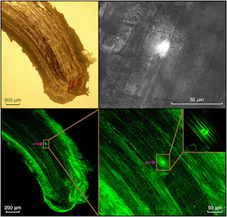

Fig. 2. Confocal laser scanning micrographs of two birch root cross sections showing the incorporation of microplastic particles in a one-year-old tree after being exposed to contaminated soil for 5 months. The location of microplastic particles are indicted by arrows and highlighted inside the circle. In the left image, the upper and right panels show the orthogonal view of the z-stacks, showing the presence of the fluorescing microplastic particle within the sample slice. The location of microplastic particles is indicated by arrows, circle, and guidelines from the circle to each z-stack. All images have been enhanced using a contrast and brightness correction. Scale is shown in the bottom right or left corner of the respsective image.
資料來源:
https://www.sciencedirect.com/science/article/pii/S0048969721071618#f0010
0 意見:
張貼留言